In the rapidly evolving world of cancer research and translational medicine, Multiplex Immunohistochemistry (mIHC) has emerged as a game-changing technology. Offering high-dimensional insights into tumor biology and immune microenvironments, mIHC is revolutionizing how researchers analyze tissue samples for clinical and scientific discovery.
This blog will explore the comprehensive suite of mIHC kits , reagents, antibodies, and technologies offered in the latest product portfolio, while optimizing content around the most relevant SEO keywords to enhance visibility on Google search results.
What Is Multiplex IHC (mIHC) ?
Multiplex IHC —also known as multiplex immunohistochemistry —is an advanced technique that allows simultaneous detection of multiple biomarkers within a single tissue section. Unlike traditional IHC, which typically labels one or two markers at a time, mIHC enables up to nine-plex analysis , providing deeper insights into cellular interactions, tumor heterogeneity, and immune cell infiltration.
✅ Key Benefits of mIHC :
- High-resolution spatial profiling.
- Simultaneous detection of multiple targets.
- Enhanced signal amplification via TSA technology.
- Compatibility with both FFPE and fresh frozen samples.
- Supports both manual and automated staining workflows.
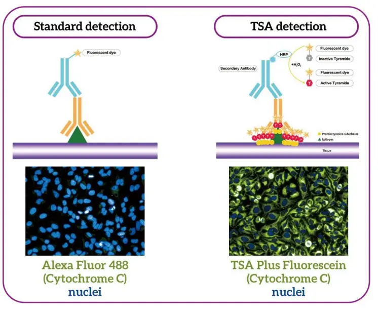
The Core Technology Behind mIHC : Tyramide Signal Amplification (TSA)
At the heart of multiplex IHC lies Tyramide Signal Amplification (TSA) technology. This powerful method significantly enhances fluorescent signal detection by depositing multiple fluorophore-labeled tyramides at antigen sites, resulting in signal amplification up to 1000x compared to conventional methods.
🔬 How TSA Works:
- Primary antibody binds to target antigen.
- Secondary antibody conjugated with horseradish peroxidase (HRP) binds to primary.
- Fluorophore-labeled tyramide is activated by HRP and covalently binds to nearby tyrosine residues.
- Multiple tyramide molecules bind near each antigen site, amplifying the fluorescent signal.
This process ensures high sensitivity , low background noise , and accurate localization of target proteins in complex tissues.
mIHC Kit Options : From 4-Plex to 7-Plex Detection
Whether you're conducting basic research or preparing for clinical trials, selecting the right mIHC kit is essential. The product line includes a wide range of options tailored for different sample types and staining methods:
🧪 Available mIHC Kits :
Kit Type | Number of Colors | Sample Compatibility | Staining Method |
|---|---|---|---|
AXT37100011 | 7-color | FFPE | Manual & Automated |
AXT36100011 | 6-color | FFPE | Manual |
AXT35100011 | 5-color | FFPE | Manual |
AXT34100011 | 4-color | FFPE | Manual |
AXT37050031F | 7-color | Fresh/Frozen | Manual |
AXT36050031F | 6-color | Fresh/Frozen | Manual + AbEraser |
Automation Kits | Up to 7-color | FFPE | Fully Automated |
These kits are available in both MSI (Manual Staining Instruments) and WSI (Whole Slide Imaging) configurations, ensuring compatibility with your lab’s workflow and imaging systems.
Key Features That Set These mIHC Kits Apart
When choosing a multiplex IHC solution , it's important to look beyond just the number of colors. Here are some standout features that make these kits ideal for advanced research:
🔑 Unique Selling Points :
- Signal Amplification : Achieve up to 1000x signal boost using TSA technology.
- Species Compatibility : Works seamlessly with mouse, rabbit, and other species-specific antibodies.
- Multi-fluorophore Detection : Supports 3–9 color combinations for deep spatial phenotyping.
- IP Protection : Proprietary formulation ensures consistent performance and reliability.
- Training Support : Access to professional operational training services for optimal use.
Comprehensive Reagent Portfolio for mIHC Workflows
To ensure reproducibility and accuracy in every experiment, the full lineup includes essential reagents for antigen retrieval, dewaxing, washing, and antibody stripping.
🧬 Required Reagents :
Part Number | Item Description |
|---|---|
AXT9510000 | HRP-labeled anti-mouse/rabbit polymer |
AXT94100001 | XTSA Antigen Retrieval Solution (pH 6) |
AXT94100002 | XTSA Antigen Retrieval for Automation (pH 9) |
AXT9710000 | AbEraser Antibody Stripping Solution |
AXTP212 | Dewax Solution |
AXTP215 | Wash Buffer (20X) |
AXTP217 | Antigen Retrieval Solution 2 |
AXTP218 | Antigen Retrieval Solution 1 |
AXTP220 | Incubation Cover Slips |
These reagents support both manual and automated protocols , making them ideal for labs transitioning from traditional IHC to high-plex mIHC.
Target-Specific Antibodies for Precision Research
The availability of validated, ready-to-use antibodies is crucial for successful mIHC experiments. The following markers have been extensively tested across various cancer types:
🧫 Targeted Biomarkers :
- Immune Markers : CD3, CD8, CD20, CD68, CD56
- Checkpoint Proteins : PD-1, PD-L1
- Cellular Markers : Ki67, PANCK
- Stromal and Follicular Markers : CD21, CD23
Each antibody comes in 12ml bottles (e.g., AXB1014-12 for CD3), ensuring sufficient supply for repeated testing.
Fluorophores : Enabling High-Dimensional Spatial Analysis
Accurate spectral separation is critical when performing multiplex IHC . The platform supports a broad spectrum of fluorophores optimized for multi-channel fluorescence microscopy.
🌈 Available Fluorophores :
Fluorophore | Excitation (nm) | Emission (nm) | Cap Color |
|---|---|---|---|
DAPI | 358 | 461 | Blue |
XTSA 480 | 436 | 485 | Purple |
XTSA 520 | 494 | 525 | Green |
XTSA 570 | 550 | 570 | Red |
XTSA 620 | 595 | 620 | Brown |
XTSA 650 | 627 | 650 | Orange |
XTSA 690 | 676 | 694 | White |
XTSA 780* | 743 | 767 | Pink |
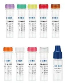
These fluorophores enable precise multicolor imaging without crosstalk, even in complex tissue architectures like tumor microenvironment mapping .
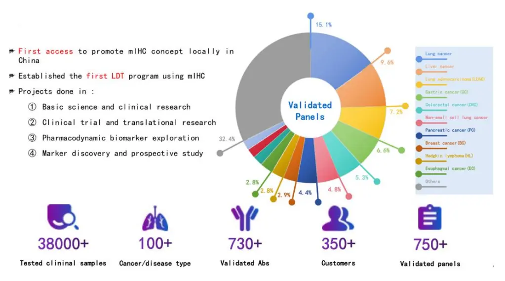
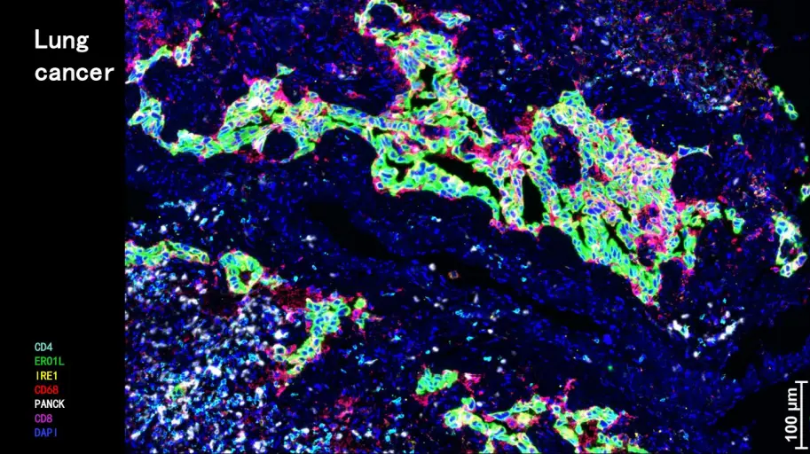
Applications Across Cancer Types and Disease Models
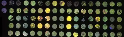
With over 38,000+ clinical samples tested , this mIHC platform has been applied across a wide range of cancers and diseases :
- Lung Cancer
- Breast Carcinoma (BCO)
- Colorectal Cancer (CRC)
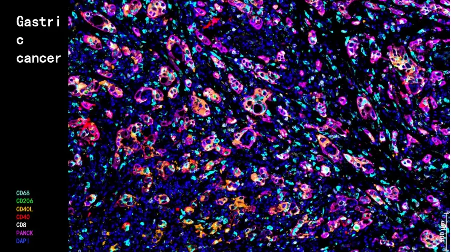
- Gastric Cancer
- Pancreatic Cancer
- Esophageal Cancer
- Hodgkin Lymphoma
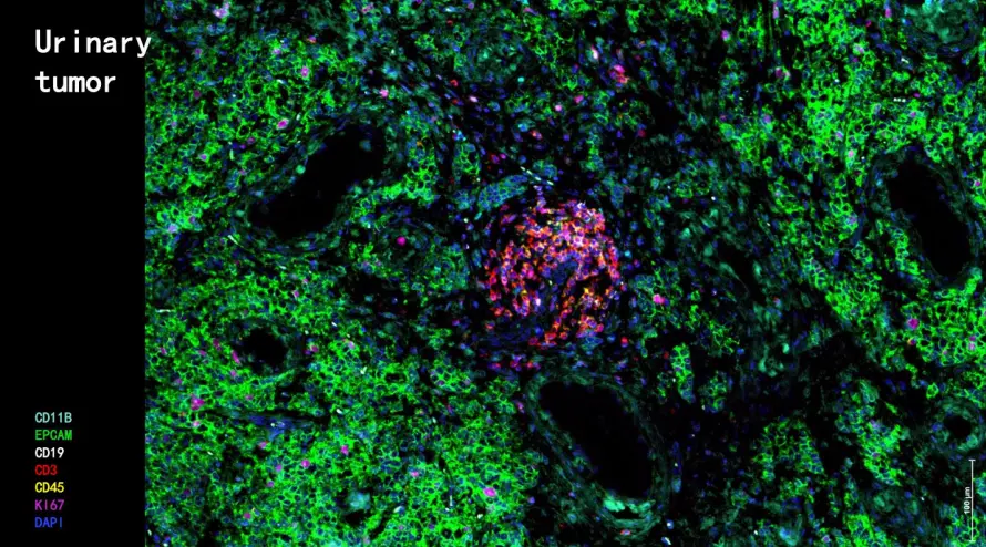
- Urinary Tumors
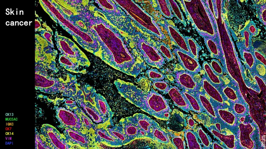
- Skin Cancer
- Liver Cancer
From marker discovery to clinical trial biomarker validation , these tools empower researchers to explore tumor-immune interactions, identify novel therapeutic targets, and monitor drug responses in real-time.
Scientific Validation Through Peer-Reviewed Publications
A robust body of peer-reviewed literature supports the effectiveness and versatility of this mIHC platform. Notable publications include :
- Nature (IF=64.8) – Liver tumour immune microenvironment subtypes
- Cancer Cell (IF=50.3) – Molecular subtypes of neuroendocrine carcinomas
- Signal Transduction and Targeted Therapy (IF=39.3) – PD-1 blockade efficacy in rectal cancer
- Kidney International (IF=19) – Single-cell analysis of parietal epithelial cells
- Clinical Cancer Research (IF=11.5) – Response to PD-1 plus chemotherapy in NSCLC
These studies demonstrate the platform’s utility in uncovering biological mechanisms , identifying prognostic signatures , and guiding immunotherapy strategies .
Why Choose This mIHC Platform?
If you’re looking for a scalable, flexible, and highly sensitive solution for spatial biology and immune profiling , this mIHC platform offers unmatched advantages :
- Proven performance across thousands of clinical samples.
- End-to-end solutions including kits, reagents, antibodies, and fluorophores.
- Customizable panels for translational research and clinical development.
- Support for both academic and industrial applications.
Multiplex IHC is no longer a luxury—it’s a necessity for modern cancer research and precision medicine. Whether you're analyzing tumor-infiltrating lymphocytes , studying immune checkpoint expression , or validating therapeutic targets , this platform equips you with the tools needed to push the boundaries of discovery.
Ready to elevate your research? Explore our full range of mIHC kits , validated antibodies , and fluorophores today—and unlock the next level of spatial biology insight.